From a pathophysiological perspective, congenital heart diseases are classified functionally, referring to the blood flow and the haemodynamic consequences of their anatomy. The advantage of this classification is its correspondence with various surgical strategies.
- Non-cyanotic heart diseases with L-to-R shunt. These present with increased pulmonary blood flow which may lead to pulmonary hypertension (Figure 14.14).
- ASD, VSD, AV canal defect
- Patent ductus arteriosus, aortopulmonary fistula
- Partial anomalous pulmonary venous return
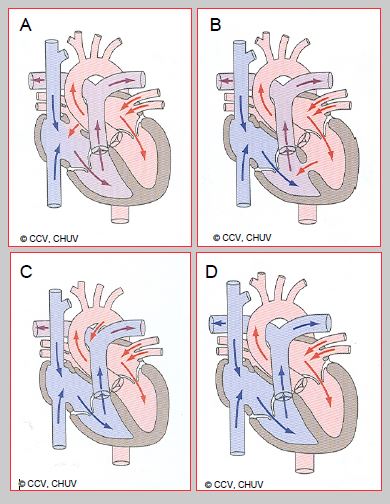
Figure 14.14: Non-cyanotic heart diseases. Non-cyanotic heart diseases with L→R shunt, which present with an increase in pulmonary blood flow, are associated with atrial septal defect (A), ventricular septal defect (B) and patent ductus arteriosus (C). In heart diseases without shunting, such as valvular heart diseases (D: mitral stenosis), the anatomical configuration of the cardiac chambers, connections, and vessels is normal. Saturation levels are normal throughout the blood flow circuit.
- Obstructive heart diseases (non-cyanotic without shunt) (Figure 14.14D).
- Mitral stenosis, cor triatriatum
- Aortic/subaortic stenosis
- Coarctation of the aorta, interrupted aortic arch
- Cyanotic heart diseases with R-to-L shunt presenting with decreased pulmonary blood flow (Figure 14.15).
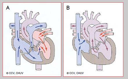
Figure 14.15: Cyanotic heart diseases with R→L shunt presenting with decreased pulmonary blood flow – pulmonary blood flow is dependent on the ductus arteriosus. A: Tetralogy of Fallot. B: Tricuspid atresia
- Cyanotic heart diseases with R-to-L shunt presenting with decreased aortic blood flow (Figure 14.16).
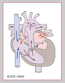
Figure 14.16: Cyanotic heart diseases with R→L shunt presenting with decreased aortic blood flow – systemic blood flow is dependent on the ductus arteriosus, as in hypoplastic left heart syndrome shown here. The ascending aorta is hypoplastic and systemic perfusion is dependent on the ductus arteriosus
- Cyanotic heart diseases with mixed or bidirectional shunt (Figure 14.17).
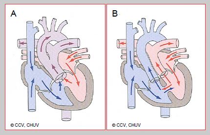
Figure 14.17: Cyanotic heart diseases with bidirectional R→L shunt. A: Truncus arteriosus. B: Transposition of the great arteries.
The age of symptom onset varies depending on lesion type. Disorders associated with low pulmonary blood flow cause early-onset cyanosis once the ductus arteriosus has closed in the first days of life. For example, children with TGA very quickly become cyanotic. Symptom onset in subjects with L-to-R shunts and high pulmonary blood flow only occurs after several weeks as PVR falls in the first two months.
© BETTEX D, BOEGLI Y, CHASSOT PG, June 2008, last update February 2020
The age of symptom onset varies depending on lesion type. Disorders associated with low pulmonary blood flow cause early-onset cyanosis once the ductus arteriosus has closed in the first days of life. For example, children with TGA very quickly become cyanotic. Symptom onset in subjects with L-to-R shunts and high pulmonary blood flow only occurs after several weeks as PVR falls in the first two months.
| Classification of congenital heart diseases |
| Pathophysiological classification - Non cyanotic L-to-R shunt - Obstruction - Cyanotic R-to-L shunt with reduced pulmonary blood flow (Qp:Qs < 1) - Cyanotic R-to-L shunt with reduced systemic blood flow (Qp:Qs > 1) - Bidirectional shunt |
© BETTEX D, BOEGLI Y, CHASSOT PG, June 2008, last update February 2020